INTRODUCCIÓN
La morfología (Anatomía) externa e interna de la abeja mellifera se corresponde esencialmente con la de los demás insectos. Lo mismo puede decirse de la fisiología (funciones vitales). No obstante Existen diferencias que es preciso indicar para una mejor comprensión de su etología (comportamiento).
Lógicamente las peculiaridades anatómicas y las funciones vitales están ínter relacionadas.
ANATOMÍA EXTERNA DE LA ABEJA
La abeja pertenece al reino animal, y dentro de él, al tipo de los artrópodos (patas articuladas), a la clase insectos himenópteros (alas membranosas) y familia de los ápidos.
El cuerpo de la abeja de la miel se divide en cabeza, tórax y abdomen, partes que están unidas y se mueven entre sí. El esqueleto externo (exoesqueleto) compuesto de quitina, que da al insecto la necesaria estabilidad, protege las tres grandes partes en que se divide el cuerpo de la abeja; en las dos primeras formando cajas rígidas y en la última de forma extensible.
El exoesqueleto, que tiene la particularidad diferencial con los vertebrados de ser externo y por lo tanto limita definitivamente el crecimiento, aloja en su interior los órganos blandos, al revés de los animales superiores, donde los órganos blandos cubren el esqueleto.
Se halla constituido por la cutícula que la forman dos capas: una exterior muy dura (exocutícula) y otra interior (endocutícula). Interiormente, el exoesqueleto se halla recubierto por la membrana basal, donde se insertan los músculos.
CABEZA
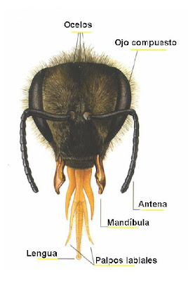
La cabeza está formada por seis escleritos íntimamente soldados entre sí.
Los ojos simples u ocelos, en número de tres, están situados en la parte superior de la cabeza, entre los ojos
compuestos, están recubiertos de pelos táctiles y tienen estructura muy sencilla.
Con ellos puede ver la abeja a corta distancia, y en condiciones de casi oscuridad en el interior de la colmena. Se ha constatado que son órganos sensibles a la intensidad de luz y son utilizados como fotómetros, determinando el principio y fin de la jornada laboral.
Los dos ojos compuestos están formados por numerosas facetas hexagonales y cada uno de ellos por miles de ojos simples (3.000 en la reina, 6.000 en la obrera y 13.000 en el zángano). La forma de las facetas hace pensar en el tipo de construcción de los panales. La visión de los colores varía con respecto a la visión humana. Tienen más agudeza visual en el lado ultravioleta del espectro. En el lado del rojo se muestran prácticamente ciegas. Ven muy bien el color azul, amarillo, verde-azulado y ultravioleta.
El color rojo lo ven como si fuera negro y dentro del amarillo confunden el naranja y el verde amarillento como si fueran amarillos.
La agudeza visual es inferior a la del hombre, pero a igualdad de tiempo, el ojo de la abeja percibe 10 veces más imágenes. Recibe la luz polarizada, la luz en la cual los rayos vibran en un solo plano.
La antena está formada por una parte rígida (escapo) y otra flexible (flagelo) se divide en segmentos (artejos). La porción que viene a continuación del "escapo" se llama pedúnculo o pedicelo, es un artejo que también forma parte del flagelo.
El número de artejos es de 12 en la reina y obrera y de 13 en el zángano.
Las antenas poseen numerosos órganos sensoriales, en forma pilosa y en placas o poros, en número de 3.000, por antena en la reina, de 3.600 a 6.000 en la obrera y unos 30.000 en el zángano, que son los responsables del tacto, oído y olfato.
Los pelos u órganos pilosos son órganos del tacto y recubren la mayor parte de la antena, y las placas o poros tienen forma de embudo y sirven para el olfato.
Si hacemos un corte transversal de la antena, y la observamos al microscopio veremos en su interior una red de nervios muy manifiestos que sirven como aparato receptor y transmisor de sensaciones.
TÓRAX
 En el tórax es donde se encuentra al aparato locomotor, estando constituido por tres segmentos o anillos,que reciben los siguientes nombres de adelante atrás: Protorax, Mesotórax y Metatórax y un pequeño segmento adicional llamado propodeo. En cada segmento lleva un par de patas, y en el segundo y tercero llevan cada uno un par de alas membranosas. También disponen de espiráculos (orificios), por donde entra el aire para la oxigenación del tórax.
Al tórax también se le llama "corselete" y en su parte superior dorsal es donde se marcan las reinas, con el color del año correspondiente según el código internacional de colores, para identificar el año de su nacimiento.
Como ya hemos visto anteriormente las abejas tienen tres pares de patas, y éstas para que puedan tener movimientos se dividen en nueve piezas llamadas artejos, dos cortos el primero de los cuales se encuentra unido al cuerpo, tres largos (el fémur, la tibia y el tarso), estando constituido este último por cuatro piezas.
El primer par de patas se encuentra situado en el protórax, y tienen una serie de dispositivos o piezas que las emplean fundamentalmente para: la limpieza de los ojos, con una especie de cepillo; dos piezas (vellum y peine o cepillo), ésta última articulada, que se cierra a voluntad para la limpieza de las antenas.
En el último artejo del tarso tiene dos garfios, que los emplean para agarrarse a superficies sobre las que quiere caminar, que pueden ser lisas o rugosas, y también para agarrarse a otras abejas, formando la llamada cadena de la cera, o cuando enjambran al formar la clásica bola o enjambre.
El segundo par de patas se encuentran situadas en el mesotórax y no tienen ninguna característica especial.
En esta parte del tórax se abre el primer par de estigmas (espiráculos), de gran importancia en el diagnóstico de la enfermedad denominada Acarapisosis.
Estas patas llevan en el extremo del tarso un garfio o espolón que emplean para desprender las pelotas de polen, que llevan en las “cestillas” del tercer par de patas.
Una especie de cepillo, la emplean para la limpieza de las alas.
 El tercer par de patas se encuentran situadas en el metatórax y son las más grandes.
Estas patas tienen los dispositivos para almacenar el polen y propóleos, llamadas corbículas o “cestillos” del polen, que se encuentran en la parte exterior de la tibia, estos cestillos tienen unos pelos fuertes y algo curvados, lo que les permite retener el polen o propóleos recogidos de las flores o de los brotes que visitan las abejas, después de ser amasado con las mandíbulas.
Los “cestillos” del polen solamente los tienen las obreras, por el contrario las reinas y zánganos carecen de ellos por no necesitarlos.
En este tercer par tienen otro dispositivo, que lo emplean a modo de pinza para recoger las
laminillas de cera elaboradas en las glándulas cereras y posteriormente pasarlas a las mandíbulas para su amasado y posterior construcción de panales.
Las alas se encuentran en el tórax, las dos primeras más grandes se insertan en el metatórax y las otras dos más pequeñas en el mesotórax.
Estos dos pares de alas están formadas por una membrana muy delgada y transparente y reforzada por una red de nervaduras quitinosas, que al mismo tiempo permiten el riego de la hemolinfa (sangre de la abeja) y el aporte de oxígeno.
Poseen nervaduras convexas y nervaduras cóncavas y tienen, en una zona determinada, una disposición
y medida (índice cubital) que sirve para clasificar las diferentes razas de abejas.
Cuando la abeja hace vuelos largos une las dos alas por medio de unos garfios o ganchos para formar una sola ala grande que hace que el vuelo sea mucho más veloz.
Por el contrario cuando hace vuelos de precisión para visitar las flores y recoger el néctar o polen estas las desenganchan y pueden quedarse quietas en el aire como las libélulas.EL ABDOMEN
El abdomen se compone de 9 segmentos, pero solo son visibles 6 en las hembras y 7 en los machos. Los segmentos abdominales poseen dos placas cada uno, llamándose a los dorsales "tergitas" y a los ventrales "esternitas", estando unidos éstos por membranas flexibles, lo que les permite una gran variedad de movimientos, como alargarse o acortarse y también curvarse en cualquier dirección.
Las membranas intersegmentarias de las esternitas, de débil consistencia, son perforadas por Varroa destructor para alimentarse con la hemolinfa de la abeja.
En cada tergita tienen un pequeño agujero que son los estigmas o espiráculos, por donde entra el aire en el interior del insecto.
El abdomen se encuentra recubierto de pelos, y según su longitud y coloración de los segmentos son índices que también se emplean para la identificación de las diferentes razas de abejas.
En el abdomen nos encontramos con: las glándulas cereras, glándula de Nosanoff y aparato de defensa.EXTERNAL ANATOMY OF THE BEE.
By: Dr. Vet. Jesus Martinez Llorente
INTRODUCTION
Morphology (anatomy) of the outer and inner mellifera bee essentially corresponds to that of other insects. The same is true of physiology (vital functions). However there differences to be indicated for a better understanding of their etiology (behavior).
Logically anatomical peculiarities and vital functions they are inter-related.
EXTERNAL ANATOMY OF BEE
The bee belongs to the animal kingdom, and within it, the kind of arthropods (jointed legs), the class Insecta Hymenoptera (membranous wings) and Apidae family.
The body of the honey bee is divided into head, thorax and abdomen, parties are united and move together. The external skeleton (exoskeleton) composed of chitin, which gives the insect the necessary stability, protects the three major parties that the body of the bee is divided; in the first two forming rigid boxes last so extensible.
The exoskeleton, which has the differential particularity to vertebrates to be external and therefore limits the growth definitely, houses inside the soft organs, unlike the higher animals, where the soft organs covering the skeleton.
It is composed of the cuticle that forms two layers: a hard outer (exocutícula) and indoor (endocutícula). Internally, the exoskeleton is covered by the basement membrane, where the muscles are inserted.
HEAD
The head, quitinosa box, which is shaped like an inverted triangle, houses the organ of vision (eyes simple and compound eyes), antennae and mouthparts. It is attached to a narrow chest and webbing of the neck.
The head consists of six escleritos closely welded together.
Simple or ocelos, three in number, eyes are located at the top of the head between the eyes
compounds, for tactile hairs are coated and have very simple structure.
With them you can see the bee at close range, and almost dark conditions inside the hive. It has been found that are sensitive to light intensity and organs are used as photometers, determining the beginning and end of the workday.
The two compound eyes are composed of numerous hexagonal facets and each by thousands of simple eyes (3,000 in queen, 6,000 and 13,000 working on the drone). The shape of the facets suggests the type of construction of the honeycombs. The color vision varies with respect to human vision. They have more visual acuity in the ultraviolet end of the spectrum. On the side of red is virtually blind. Look great blue, yellow, green-blue and ultraviolet.
The red color they see like black and inside yellow confuse orange and yellow-green as if they were yellow.
Visual acuity is lower than that of men, but equal time, the bee eye perceives images 10 times. Receive polarized light, in which light rays vibrate in a single plane.
The two antennas emerge from the center of the face, being closely spaced articulating head through a membrane.
The antenna consists of a rigid part (scape) and other flexible (flagellum) is divided into segments (knuckles). The portion that follows the "escaped" is called stem or stalk, is a knuckle which is also part of the flagellum.
Artejos number is 12 in the queen and workers and 13 in the drone.
The antennas have many sensory organs, hairy and shaped plates or pores, numbering 3,000, for antenna Queen of 3600-6000 in the working and about 30,000 in the drone, which are responsible for touch, hearing and smell.
The hairs or hair organs are organs of touch and lining most of the antenna, and the plates or funnel-shaped pores and are used to smell.
If we make a cross section of the antenna, and observed to see inside very manifest a network of nerves that serve as transmitter and receiver apparatus sensations microscope.
CHEST
In the chest is where the locomotor system, being constituted by three segments or rings, which are named as follows front to back: Prothorax, Mesothorax and Metathorax and an additional segment called propodeum. In each segment it carries a pair of legs, and the second and third each carry a pair of membranous wings. They also have spiracles (openings), through which enters the air for oxygenation of the chest.
The chest is also called "thorax" and its top ridge is where the queens are marked with corresponding color year according to the international color code to identify the year of his birth.
As we have seen before bees have three pairs of legs, and so they can have these movements are divided into nine parts called knuckles, two short the first of which is attached to the body, three short (femur, tibia and Tarsus), the latter being composed of four parts.
The first pair of legs is located on the prothorax, and have a number of devices or parts that primarily used for: cleaning the eyes, a sort of brush; two pieces (vellum and comb or brush), the latter being articulated, which closes at will for cleaning antennas.
In the last knuckle of Tarsus has two hooks, that used to cling to surfaces on which you want to walk, which can be smooth or rough, and also to hold on to other bees, forming the so-called chain of wax, or when the swarm forming the classic ball or swarm.
The second pair of legs are located on the mesothorax and have no special feature.
In this part of the chest the first pair of stigmata (spiracles) opens, of great importance in the diagnosis of the disease called Acarapisosis.
These legs take on the end of a hook or tarsal spur used to dislodge the balls of pollen, leading into the "baskets" of the third pair of legs.
A kind of brush, used for cleaning the wings.
The third pair of legs are located on the metatórax and are the largest.
These legs have devices to store pollen and propolis, called corbiculae or "buckets" pollen found in the outside of the tibia, these buckets have strong hairs and somewhat curved, allowing them to retain pollen or propolis collected from the flowers or buds visiting bees, after being kneaded with the jaws.
The "baskets" of pollen only have the workers, by contrast queens and drones lack them not to need them.
In this third pair they have another device, employing the pincer to collect
wax flakes made in the wax glands and then pass them to the jaws for kneading and subsequent construction of honeycombs.
The wings are located in the thorax, the first larger insert into two metathorax and two smaller on the mesothorax.
These two pairs of wings are formed of a thin, transparent membrane and reinforced by a network of chitinous ribs at the same time allow the irrigation of the hemolymph (blood of the bee) and oxygen.
Have convex and concave ribs and ribs have, in a certain area, a provision
and measurement (cubital index) used to classify the different races of bees.
When the bee makes long flights linking the two wings by means of hooks or hooks to form a single large wing that makes flying much faster.
On the contrary when you visit precision flying flowers and collect nectar or pollen these the disengaged and can stand still in the air like dragonflies.
THE ABDOMEN
The abdomen consists of 9 segments, but are visible only 6 females and 7 males. Abdominal segments each possess two plates, calling dorsal "tergitas" and ventral "sternites", these being connected by flexible membranes, which allows a variety of movements, such as extended or shortened and bent in any direction .
Intersegmental membranes sternites of weak consistency, being pierced by destructive feeding with Varroa bee hemolymph.
In each tergita they have a small hole which are stigmas or spiracles, where it enters the air inside the insect.
The abdomen is covered with hairs and along its length and color indexes of the segments are also used for the identification of different strains of bees.
In the abdomen we find: the wax glands, gland Nosanoff and defense apparatus.


No hay comentarios:
Publicar un comentario
Gracias - Thank you.